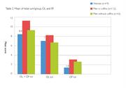
Figure 3. Total mean score of osteolytic lesions and osteophytic formation/group.
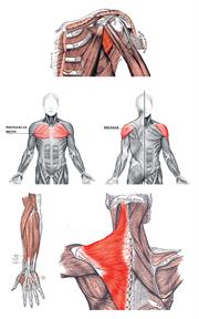
Figure 4. From left to right, upper row: the subscapularis, the pectoralis major and the deltoid muscles.
Lower row: the extensors and the trapezium muscle (from the 20th U.S. edition of Gray's Anatomy of the Human Body, originally published in 1918). Source: Wikimedia Commons.
The treatment of the material in this way is justified by several differences between the burials in question. Both the burial customs (see above) and strontium isotope the analysis values put the skeletons neatly into two separate groups. It is possible to link the strontium isotopes values to the written sources, which tell of war prisoners that were forced to work in the mine as a punishment during the 16th and 17th centuries. Most likely, the men buried without coffins represent soldiers from Poland, Germany, Russia or Denmark. The idea is that they worked in the mine for a shorter period of time, and thus did not obtain “typical lesions attributed to miners”. They are not “real miners”. Hypothetically, this group should then display a different pattern of enthesopathies of the skeletons compared to the men buried in coffins.
The bar chart (Figure 3), shows that the total mean score of pathological changes of the muscle attachments are slightly higher in the group of men buried in coffins. Does this mean that they have had a heavier workload? The fact that the men buried without coffins probably had a different ethnic origin in comparison with the men, women, infants and juveniles buried in coffins could also influence the development of skeletal lesions, as well as occupational status.
Provided the women did the same type of labour in the mine as the men, we would expect the scores for women to be approximately the same as the scores for “the real-miners”. The bar chart shows that the total mean score for enthesopathies in total for women is lower than the two groups of men, although the total mean scores of porosity (OL) are slightly higher for women than for men buried without coffins (Figure 3). The group of women are in general older than this group of men and should therefore, if we consider the correlation between older ages and more lesions, display a higher mean score, but then we have not accounted for the probable correlation between sex and lesions. Anyway, the women display more similarities to the group of “non-miners” and therefore they probably performed other tasks than the so called “real-miners”. What kind of tasks did they perform? Are they consistent with the information we get from the written contemporary sources?
To get a grip of these questions I have narrowed the survey to only encompass women and men buried in coffins. To do a more detailed study of differences and similarities in the pathological changes of the different muscle attachments, the humerus and the clavicle have been selected, i.e. the bones exhibiting the highest total scores of enthesopathies in both sexes. Porosity (OL) and bone formation (OF) were analyzed separately on 10 muscle attachments on the humerus and three on the clavicle.
In fact, there are some differences seen between the sexes in the attachments affected in each of the bones. Almost half of the women show “repetitive strain injuries” (both porosity and bone formation) on the subscapularis muscle on humerus, a muscle that medially rotates the arm at the shoulder (Figure 4). Some 73 percent of the women display strain (porosity) on the clavicle’s deltoid muscle. The deltoid muscle flexes, medially rotates and moves the arm away from the body at the shoulder.
Bone formation (OF) can be seen in c. 18 percent of the women on the trapezius muscle on the clavicle. This muscle acts on the shoulder: it elevates and upwardly rotates the shoulder blade and extends the neck. It also retracts the shoulder blade.
The men on the other hand seem to have used the pectoralis major muscle on the humerus to a great extent. Over half of the men show strain on this muscle (porosity). The pectoralis maior flexes, extends, medially rotates and adducts the arm at shoulder. On the clavicle the deltoid muscle displays strain (porosity) among c. 46 percent of the men.
Also, c. 40 percent of the male skeletons display bone formations (OF) on the extensors and the extensor carpi radialis longus on humerus. The extensors extend wrist and fingers, extensor carpi radialis longus extends and radial deviates (abducts) the hand at the wrist.