Table 1.
How do bodies, or in this case skeletons, adjust to different work load? Is it possible from the skeletons to draw conclusions about how women and men used their bodies, and is there, in this case-study, any indication of women working in the mine? In the following article I will discuss work from a physiological point of view, focusing on the skeletons buried at Sala mining cemetery and especially the skeletons of women.
There are different physiological and biological properties inherent in the skeletons of women and men respectively. In general, the skeletons of men are more robust and exhibit more marked attachments for muscles and ligaments of the bones. This reflects the fact that men possess a larger muscle mass. This suggests heavier duties for men, a view that often is stressed in written sources and contemporary literature, for instance the work in mines.
We know mining in Europe becomes more and more looked upon as a predominantly male occupation during the second half of the 19th century . But what did the gender division look like during the 16th century when the “proto-industry” of mining in Sweden started to become more of a concern to the Crown.
Statement/Hypothesis: Mining below ground is regarded as a heavy and dangerous work that ought to cause stress lesions on the skeleton. In what ways have the skeletons of the women buried in the mining cemetery been affected, and can the lesions on the muscle attachments be explained in terms of labour division?
The following variable will be used: Pathological changes of the attachment sites for muscles, i.e. pitting and/or new bone formation
The use of enthesopathies, i.e. pathological changes of the entheses, to distinguish between different types of work or to discuss gender divisions of labour have been questioned by several researchers. One problem is the correlation between older ages and presence of enthesopathies, and also a probable correlation between sex and enthesopathies. Other problems are the range of different methods available (not good for comparisons), and also methods that are not sufficiently stringent, which increases the risk of being subjective and making incorrect assessments of the lesions.
Additional information about the “use of bodies” can be gained from changes in larger joints and in the spine, tooth wear connected to specific occupational habits, biomechanical properties of bone, fractures and so forth. Ideally, all of these variables should be incorporated in an investigation about “manifestations of work” in skeletons.
This paper therefore offers a brief osteological perspective on gender divisions of work rather than a thorough investigation and evaluation of osteological methods or a presentation of results. The study’s small sample size must also be taken into account.
The mining cemetery
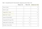
Parts of the cemetery, situated in the county of Västmanland in the middle of Sweden, were excavated in the beginning of the 2000s. The cemetery seems to be divided into two areas differing regarding burial customs, sex and age. Burials without coffins are concentrated in the southern part of the excavated area, coffin burials are found in the northern part and to the south of the presumed church or chapel. Men, and preferentially younger men, are buried without coffins. Buried in coffins are members of families (women, men and children of different ages). In total, 102 skeletons were analysed osteologically: 13 women, 37 men and 52 infants and juveniles (Table 1).
The people buried in the cemetery lived in the mining village situated directly to the south of the silver mine. Excavations of the village during the 1950s and 80s date the start of the settlement to approximately the second half of the 15th century. In the beginning of the 17th century the villagers were forced by the Crown to abandon the village when Sala city, c. 4 km to the north-east of the village, was founded.